#Imagej invert colors update
same as ImageJ, but comes with many pre-installed plugins and can update itself automatically. For the Cell Counting Kit-8 (CCK-8, Dojindo, Shanghai, China) assay, 10 μL CCK-8 medium was added to each well at 0, 24 or 48 h after transfection. Cells were seeded on a 96-well culture plate with 4000 cells per well in 100 μL of culture medium and left to grow for 12 h. Positive VZ cells were identified by the presence of a BrdU-labeled nucleus that was negative for Hu staining and was touching the ventral ventricular surface above HVC (see Fig. Found insideThis book provides the first comprehensive account of multilineage-differentiating stress-enduring (Muse) cells, a pluripotent and non-tumorigenic subpopulation of mesenchymal stem cells (MSCs) that have the ability to detect damage signals. Any solutions or suggestions would be greatly appreciated However, I have a hard time removing the overlap between cells and for the program to distinguish between the clumps.

#Imagej invert colors software
αSMA, and FSP1 staining using NIHI ImageJ software in each group. CD11b single positive cells in both ischemic core (E-E') and border zone (where Ym1 is not expressed, F-F') surround neurons. It is possible to count with an additional channel (ex. plot ( x, y, marker = 'o', color = 'r', markersize = 2, markeredgewidth = 0.3.3. distance_transform_edt ( bw_blobs ) # Get ultimate points y, x = approx_ultimate_points ( bw_blobs, bw_dist ) # Show results fig = create_figure ( figsize = ( 8, 4 )) show_image ( im_blobs, title = "(A) Original image", pos = 141 ) show_image ( bw_blobs, title = "(B) Thresholded image", pos = 142 ) show_image ( bw_dist, title = "(C) Distance transform", pos = 143 ) show_image ( im_blobs, title = "(D) Ultimate points", pos = 144 ) plt.
#Imagej invert colors zip
center_of_mass ( lab_ultimate, lab_ultimate, index = range ( 1, n + 1 )) return zip ( * com ) # Load images im_blobs, bw_blobs = read_blobs_and_threshold () # Create distance transform image bw_dist = ndimage. bitwise_and ( bw, bw_maxima ) # Reduce all ultimate point regions to a single point # (Here, we take the centroids - this wouldn't be robust for weird shapes with holes, # but should be ok here) lab_ultimate, n = ndimage. binary_dilation ( bw_maxima ) bw_ultimate = np. h_maxima ( bw_dist, 0.5 ) bw_maxima = ndimage. distance_transform_edt ( bw ) from skimage.morphology import extrema bw_maxima = extrema. """ if bw_dist is None : bw_dist = ndimage. Much as I'd like to eliminate this step & just use an ImageJ screenshot, I've included it to admit the awkwardness of image processing - often methods need to be adapted to overcome seemingly minor issues. To approximate the results here, we can use h-maxima (maxima greater than a fixed threshold) with a small dilation to merge maxima that are nearby. ImageJ's 'MaximumFinder' does *a lot* or work, and so generally ImageJ will give a reasonable result without the user needing to know what exactly happened. However, in practice this can detect lots of spurious peaks / saddle-points.


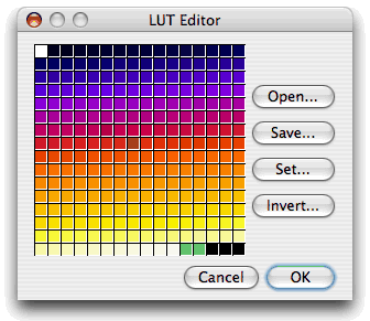
This *sounds* easy: check where pixel values match the result of applying a maximum filter, and those are your maxima. These are local maxima that are non-zero in the binary image. mean () return im_blobs, bw_blobs def approx_ultimate_points ( bw, bw_dist = None ): """ Find the ultimate points from a distance map of a binary image. """ # Load & invert blobs (this compensates for the inverted LUT) im_blobs = 255 - load_image ( 'blobs.gif' ) # Threshold blobs - we don't need to be sophisticated, # almost any half-sensible threshold method should work bw_blobs = im_blobs > im_blobs. """ def read_blobs_and_threshold (): """ Read the blobs.gif image & convert to binary in a 'standardized' way, for consistency across figures. The results aren't guaranteed to be identical, but should be close - and I hope more useful for learning. """ Rather than simply paste screenshots from ImageJ, here I've tried to use Python/skimage/scipy to replicate the steps taken within ImageJ.


 0 kommentar(er)
